Quick Facts
- Primary Cause: Chronic GERD is responsible for approximately 57% to 66% of all diagnosed cases.
- Key Symptoms: Chest pain is reported in 71.8% of patients, followed closely by painful swallowing (odynophagia) in 70% of cases.
- Incidence Rate: Drug-induced esophageal injury occurs at an estimated annual rate of 3.9 cases per 100,000 individuals.
- Diagnosis: The gold standard for identifying an ulcer is an EGD procedure (endoscopy).
- Warning Signs: Unexplained weight loss of ≥10 lbs or signs of internal bleeding like vomiting blood are critical red flags.
- Healing Timeline: With proper acid suppression therapy, most ulcers heal within a 4 to 8 week window.
An esophageal ulcer is a necrotic defect in the esophageal mucosa that penetrates into the submucosal layer, often triggered by chronic GERD or NSAID use. Unlike surface-level erosion, an esophageal ulcer involves deeper tissue damage that requires specific medical intervention to prevent serious complications like hemorrhage or strictures.
Recognizing Esophageal Ulcer Symptoms and Warning Signs
When you experience persistent discomfort in your chest, it is easy to dismiss it as standard acid reflux. However, distinguishing between common heartburn and the more severe esophageal ulcer symptoms is vital for your long-term digestive health. While heartburn often feels like a rising, burning sensation—frequently illustrated by a vertical hand motion moving up the chest—ulcer pain tends to be more localized and intense.
The hallmark of an ulcerated esophagus is often odynophagia, or painful swallowing. This is not just a "scratchy throat" sensation; it is a sharp, stabbing pain felt deep in the chest when food or liquid passes the ulcerated site. Many patients also struggle with dysphagia, the sensation of food getting stuck, which occurs as the inflammation disrupts the normal rhythmic contractions of the esophagus.
We also look for specific "alarm symptoms" that suggest the condition is progressing. If you notice a significant drop in energy alongside shortness of breath, it may indicate chronic blood loss leading to iron-deficiency anemia, often defined clinically by a ferritin level ≤45 ng/mL. Even more urgent are signs of hematemesis, which is the medical term for vomiting blood or material that looks like coffee grounds.
| Feature | Standard Heartburn | Esophageal Ulcer Warning Signs |
|---|---|---|
| Pain Type | Diffuse burning sensation | Localized, sharp chest pain |
| Swallowing | Usually normal | Painful (Odynophagia) or difficult (Dysphagia) |
| Weight Changes | None or related to diet | Unexplained weight loss ≥10 lbs |
| Bleeding Signs | None | Vomiting blood or black, tarry stools |
| Response to Antacids | Often immediate relief | Temporary or minimal relief |

Primary Causes of Esophageal Ulcers: Distal vs. Mid-Esophagus
Understanding the causes of esophageal ulcers often starts with identifying where the damage is located. In the world of gastroenterology, the "where" usually tells us the "why."
The majority of ulcers occur in the distal esophagus, which is the lower portion near the stomach. This area is the primary battleground for GERD and esophageal ulcers. When the lower esophageal sphincter fails to close properly, the esophageal mucosa is repeatedly bathed in gastric acid and pepsin. This caustic environment eventually erodes the lining, leading to a necrotic defect. Research indicates that gastroesophageal reflux disease remains the leading driver of these injuries.
In contrast, ulcers found in the mid-esophagus are frequently "pill-induced." This happens when certain medications, particularly NSAIDs or antibiotics like doxycycline, get physically stuck or linger too long in the mid-portion of the tube. These medications can cause direct chemical irritation to the tissue. Additionally, for those with weakened immune systems, infections such as Candida albicans can take hold, creating diffuse ulcerations throughout the esophageal lining.
| Ulcer Location | Primary Cause | Typical Trigger |
|---|---|---|
| Distal (Lower) | Acid Reflux | Chronic GERD, Hiatal hernia |
| Mid-Esophagus | Medication / Infection | NSAIDs, Doxycycline, Candida, or Herpes |

There are several risk factors for developing esophageal ulcers that you should keep on your radar. Tobacco use and excessive alcohol consumption both weaken the esophageal lining and increase acid production. Furthermore, the habit of taking medication without enough water or lying down immediately after swallowing a pill significantly increases the risk of localized chemical burns.
Diagnosis: The EGD Procedure and Biopsy Protocols
If your symptoms persist or if you fall into a higher-risk category—such as being age ≥60 with new-onset digestive issues—your doctor will likely recommend an EGD procedure for esophageal ulcer diagnosis. During this procedure, a gastroenterologist uses a thin, flexible tube with a camera to visualize the esophageal mucosa directly.
This visualization is critical because it allows the specialist to see the depth of the defect. We are looking for more than just surface redness; we are looking for a clear break in the tissue that extends into the submucosal layer. During the EGD, the physician will also take biopsies. These tissue samples are essential for ruling out malignancy and identifying specific infections. For instance, if a viral infection is suspected, the doctor will take samples from the center of the ulcer to check for Cytomegalovirus (CMV), or from the outer edges to look for Herpes Simplex Virus (HSV).
Another vital part of the diagnosis is screening for Barrett’s esophagus. This is a condition where the normal lining of the esophagus changes to resemble the lining of the intestine, usually due to long-term acid exposure. Identifying this early is a priority, as it is a known precursor to esophageal cancer.
Potential Complications: From Strictures to Barrett’s
Ignoring an esophageal ulcer is not just about enduring pain; it is about avoiding long-term structural damage. One of the most common signs of esophageal ulcer complications is the development of peptic strictures. These are areas of scar tissue that narrow the esophagus, occurring in roughly 60% to 70% of patients with chronic untreated reflux. If you find that you have to chew your food into a liquid consistency just to get it down, a stricture may already be forming.
Beyond narrowing, there is the risk of perforation, where the ulcer eats completely through the esophageal wall. This is a medical emergency that can lead to severe infection in the chest cavity. There is also the progression risk to Barrett’s esophagus, which affects approximately 6-12% of patients with chronic GERD. Because these cells are abnormal, they carry a higher risk of transforming into adenocarcinoma, a type of cancer. This is why we emphasize that "persistent heartburn" is a symptom that deserves a professional evaluation.
Managing Pain and Promoting Healing
The good news is that the esophagus is a remarkably resilient organ if given the right environment to heal. The primary goal of treatment is to reduce acid production to allow the mucosa to regenerate.
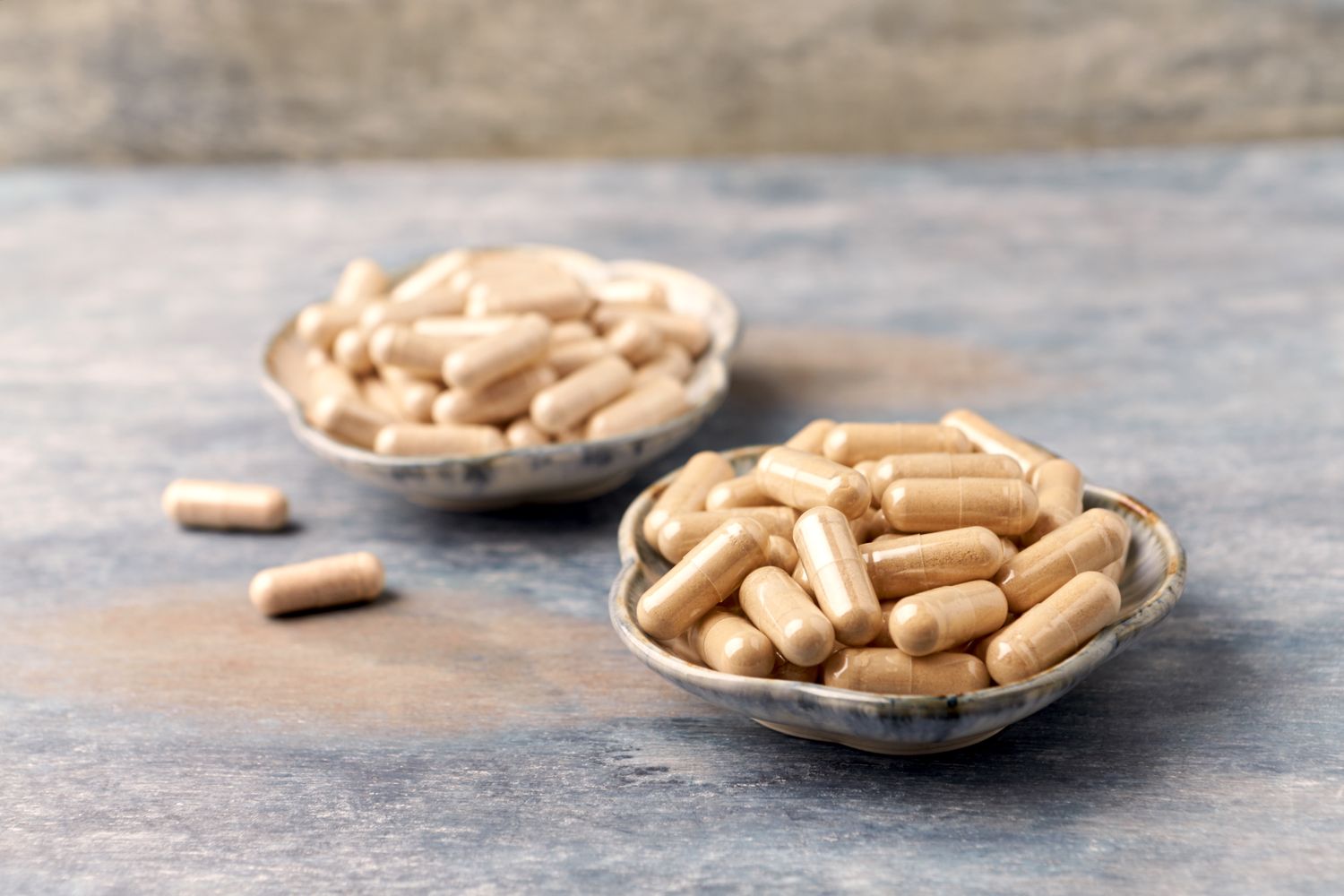
- Proton Pump Inhibitors (PPIs): These are the heavy hitters of treatment, effectively shutting down the pumps that produce stomach acid.
- H2-Receptor Antagonists: Often used for milder cases or as a secondary support, these help block the signals that trigger acid production.
- Sucralfate: Sometimes prescribed as a "liquid bandage," this medication coats the ulcer site to protect it from further irritation while you eat.
When it comes to managing esophageal ulcer pain at home, your diet plays a supporting role. We recommend avoiding "the big triggers": spicy foods, highly acidic citrus, caffeine, and alcohol. These substances can irritate the raw ulcerated tissue and delay the healing process. Smoking cessation is also non-negotiable, as nicotine relaxes the lower esophageal sphincter, allowing more acid to splash upward.
Most patients find that with consistent use of Proton pump inhibitors and lifestyle adjustments, the tissue begins to heal within 4 weeks, with full resolution typically occurring by the 8-week mark.
FAQ
What are the most common symptoms of an esophageal ulcer?
The most frequent symptoms include localized chest pain, which affects about 71.8% of patients, and painful swallowing (odynophagia), reported by 70%. Many people also experience persistent heartburn, a feeling of food being stuck in the throat (dysphagia), and occasionally nausea or a sour taste in the mouth.
What is the main cause of an ulcer in the esophagus?
The leading cause is chronic gastroesophageal reflux disease (GERD), which accounts for 57% to 66% of cases. Other significant causes include the use of certain medications like NSAIDs or antibiotics that cause "pill-induced" injury, and infections like Candida or Herpes, especially in individuals with compromised immune systems.
How long does it take for an esophageal ulcer to heal?
With appropriate medical treatment, specifically acid suppression therapy using PPIs, most esophageal ulcers heal within 4 to 8 weeks. Recovery time can vary depending on the severity of the ulcer and how strictly the patient follows dietary and lifestyle recommendations.
Is an esophageal ulcer a serious condition?
Yes, it is a serious medical condition because it involves a deep defect in the esophageal lining. If left untreated, it can lead to dangerous complications such as internal bleeding (hemorrhage), the formation of strictures that make swallowing difficult, or a perforation in the esophagus.
How do doctors diagnose an esophageal ulcer?
The primary method of diagnosis is an EGD procedure (upper endoscopy). This allows a gastroenterologist to see the ulcer directly, assess its depth, and take tissue biopsies to rule out cancer or identify specific infections that might be causing the ulcer.